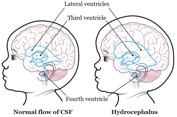
Hydrocephalus does not have a cure. The only way to treat Hydrocephalus is through brain surgery. A patient with hydrocephalus may require multiple brain surgieries in their lifetime
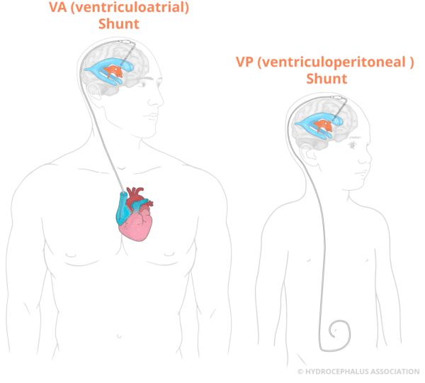
Shunt systems manage the flow of excess cerebrospinal fluid (CSF) in hydrocephalus patients. They consist of a catheter placed in the brain, a valve to regulate fluid flow, and another catheter that directs fluid to the abdomen for absorption. Shunts help maintain normal brain pressure, relieving symptoms and preventing complications. While durable, they may require adjustments or replacements over time.

VP (ventriculoperitoneal) shunts redirect excess fluid from the brain's ventricles into the peritoneal cavity, located in the abdomen, where the digestive organs reside.
VA (ventriculoatrial) shunts channel fluid from the brain to the heart, with the distal catheter placed into a vein in the neck and advancing the fluid into the right atrium.
VPL (ventriculopleural) shunts divert excess fluid into the pleural cavity, a space between the chest wall and lungs lined by membranes that facilitate fluid absorption.

VP (ventriculoperitoneal) shunts redirect excess fluid from the brain's ventricles into the peritoneal cavity, located in the abdomen, where the digestive organs reside.

The traditional procedure involves creating an opening in the floor of the third ventricle using an endoscope and a small surgical instrument. This method is typically used for patients with obstructive hydrocephalus due to aqueductal stenosis or other blockages in the ventricles.
This procedure is primarily used in pediatric patients, especially infants. Along with creating an opening in the third ventricle, the surgeon also cauterizes (burns) parts of the choroid plexus, the tissue responsible for producing CSF. Reducing CSF production can further help alleviate symptoms and is often paired with ETV in young children who might otherwise require a shunt.
In some cases, the initial ETV may close or become ineffective over time. A revisional ETV can be performed to reopen or modify the original pathway for CSF drainage. This is typically required if the symptoms of hydrocephalus return.
In cases where a brain tumor is causing the blockage, ETV can be combined with tumor resection surgery. The surgeon uses the endoscope to remove part of the tumor while also creating a pathway for fluid drainage, potentially treating both the tumor and hydrocephalus at the same time.
